
File:Siphovirus.tiff

此TIF文件的JPG预览的大小:594 × 599像素。 其他分辨率:238 × 240像素 | 476 × 480像素 | 761 × 768像素 | 1,048 × 1,057像素。
原始文件 (1,048 × 1,057像素,文件大小:1.09 MB,MIME类型:image/tiff)
摘要
| 描述 |
English: Electron micrographs of bacteriophages from Cutibacterium acnes (formerly Propionibacterium acnes). Phages were negatively stained with 0.75% uranyl formate and subjected to transmission electron microscopy. The phages have a head of approximately 55 nm in diameter, loaded with genetic material. Their tails have a size of 150 × 10 nm and are flexible and non-contractile. The upper micrographs show Propionibacterium phage PAD40 and PAD11 (NBBI TaxIDs 504513 and 504513 respectively). |
| 日期 | |
| 来源 | BMC Microbiology 2008, 8:139 doi:10.1186/1471-2180-8-139 |
| 作者 | Rolf Lood, Matthias Mörgelin, Anna Holmberg, Magnus Rasmussen and Mattias Collin |
许可协议
文件历史
点击某个日期/时间查看对应时刻的文件。
| 日期/时间 | 缩略图 | 大小 | 用户 | 备注 | |
|---|---|---|---|---|---|
| 当前 | 2011年12月2日 (五) 14:31 | 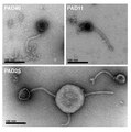 | 1,048 × 1,057(1.09 MB) | Alexbateman |
文件用途
以下页面使用本文件:
全域文件用途
以下其他wiki使用此文件:
- ar.wikipedia.org上的用途
- arz.wikipedia.org上的用途
- ca.wikipedia.org上的用途
- de.wikipedia.org上的用途
- en.wikipedia.org上的用途
- it.wikipedia.org上的用途
- ja.wikipedia.org上的用途
- www.mediawiki.org上的用途
- www.wikidata.org上的用途